Wednesday, 25 July 2018

Tools in Bioinformatics


Bioinformatics is the theory, application, and development of computing tools to solve problems and create hypotheses in all areas of biological sciences. Biology in the post-genome world has been and continues to be transformed from a largely laboratory-based science to one that integrates experimental and information science. Bioinformatics has contributed to advances in biology by providing tools that handle data sets too large and/or complex for manual analysis. Examples of some of these tools include assembling the DNA sequence of entire genomes, gene finding algorithms, microarray expression analysis, molecular systems modeling, and biomarker discovery from mass spectra. Computational tools are central to the organization, analysis, and harvesting of biological data at the level of macromolecules, cells, and systems. Consequently, there is a growing need for trained professionals who understand the languages of biology and computer science. Biologists trained in more traditional programs may not have a working knowledge of statistics and algorithms, whereas computer scientists trained in more traditional programs may not have a working knowledge of the chemistry and biology required in the field.

The Undergraduate Bioinformatics Degree at the University of Pittsburgh, which is operated jointly by the Departments of Biological Sciences and Computer Science, the program offers training that builds a solid foundation in chemistry, biology, computer science, mathematics, and statistics. This training will enable students to communicate fluently with experts across these disciplines and to have the skills necessary to apply computing tools to address contemporary problems in biology and medicine. The training will enhance the professional opportunities for undergraduates to pursue careers in pure or applied research in academia, government, pharmaceutical, medical, or biotechnology sectors.

Tuesday, 24 July 2018

Proteome of the Human Heart Mapped for the First Time


A healthy heart beats about two billion times during a lifetime – thanks to the interplay of more than 10,000 proteins. Researchers from the Max Planck Institute of Biochemistry (MPIB) and the German Heart Centre at the Technical University of Munich (TUM) have now determined which and how many individual proteins are present in each type of cell that occurs in the heart. In doing so, they compiled the first atlas of the healthy human heart, known as the cardiac proteome. The atlas will make it easier to identify differences between healthy and diseased hearts in future.

Proteins are the molecular machines of cells, in which they perform a range of functions. They are produced by the cells based on blueprints stored in their DNA. Changes occurring at the DNA or protein level can lead to disorders. For such changes to be recognized as underlying causes of heart disease, it is important to know precisely which proteins are present in the healthy heart and in what quantities.

PROTEIN MAP OF THE HEART


The first such protein atlas of the heart was recently published in “Nature Communications” by a research team from Munich. The scientists determined the protein profile of cells in all the regions of the heart, such as heart valves, cardiac chambers, and major blood vessels. In addition, they investigated the protein composition in three different cell types of the heart: cardiac fibroblasts, smooth muscle cells, and endothelial cells. In this way, the researchers were able to map the distribution of proteins in the various regions of the heart. Using mass spectrometry, they identified nearly 11,000 different proteins throughout the heart. Previous studies had focused for the most part only on individual cell types, or they used tissue from diseased hearts. “This approach has two problems,” says Sophia Doll of the MPIB and lead author of the study. “First, the results did not give a full picture of the heart across all its regions and tissues; and second, comparative data on healthy hearts were often missing. Our study has eliminated both problems. Now the data can be used as a reference for future studies.”

“Looking at the protein atlas of the human heart, you can see that all healthy hearts work in a very similar manner. We measured similar protein compositions in all the regions with few differences between them,” says Sophia Doll. We were also surprised to find that the right and left halves of the heart are similar, despite having quite different functions: the right half pumps oxygen-poor blood to the lungs, while the left half pumps oxygen-rich blood from the lungs to the body.

SICK VS HEALTHY: IDENTIFYING DIFFERENCES


In the next step, the team wanted to test whether the data from healthy hearts could serve as a control for detecting changes in diseased hearts. They compared their values with the cardiac proteomes of patients with atrial fibrillation, a very common rhythm disorder of the heart. The results indeed provided initial clues as to the cause of the disease. The tissue of the diseased hearts was most different in proteins responsible for supplying energy to the cells.

The comparison provided yet another interesting finding: Although the proteins involved in energy metabolism were changed in all the patients, those changes differed between individuals. “These findings show us how important personalized medicine is. Although all the patients had very similar symptoms, we see from the data that a different molecular dysfunction was responsible in each case. We need to learn to recognize and treat such individual differences − especially in cardiac medicine,” says Adjunct Teaching Professor Dr. Markus Krane, Deputy Director of the Department of Cardiovascular Surgery of the German Heart Centre Munich at TUM.

NEARLY 11,000 PROTEINS IN LESS THAN TWO DAYS


Together with his colleagues at the Department of Cardiovascular Surgery (Director: Professor Rüdiger Lange), Markus Krane has collected more than 150 tissue samples from over 60 cardiac operations and forensic samples. Using elaborate cell culture methods, they were able to extract the various cell types from them. This large amount of cardiac material was a crucial factor for studying the individual heart regions so precisely. Professor Matthias Mann, director of the Department of Proteomics and Signal Transduction at the MPIB, and his team carried out extensive mass spectrometric measurements. Thanks to advances in mass spectrometry and sample processing, the researchers are lighting the way towards personalized medicine. The team at MPIB attaches great importance to precise, repeatable and fast analytical methods. They have improved the measuring technique to the extent that an entire heart region can now be determined in less than two days − twice as fast as before. This is crucial, especially for potential use on patients.

Reference:https://www.technologynetworks.com/proteomics/news/proteome-of-the-human-heart-mapped-for-the-first-time-294214

Monday, 23 July 2018

Future of the World Bioinformatics

Bioinformatics is one of the software tools for understanding the biological data. Sequence analysis is the process of regulating the DNA and RNA sequence in the analytical method for understanding its features, functions, structures, and evolution. It determines the sequence of the polymer formed of several monomers and revealing the evolution and genetic diversity of sequences and organisms. Gene expression is the most fundamental level at which genotype gives rise to the phenotype. The amount of gene expression can have a deep effect on the functions of the gene in a cell or in a multicellular organism. The bioinformatics workflow management system is specifically designed to compose and execute a series of manipulation steps that are related to bioinformatics. Bio Computer objects intents to facilitate bioinformatic workflow related exchange and communicate between the regulatory agencies and pharmaceutical companies and researchers.

To know more about it visit the website of the conference 


Wednesday, 18 July 2018

Industrializing Proteomics

Industrializing Proteomics to Transform the Future of Healthcare


Researchers are discovering a plethora of potential new biomarkers every year, each touted as the ‘next big thing’ that will help herald a new era of precision medicine. But so far, very few have made it into clinical practice. We find out how some proteomics laboratories are now tackling this bottleneck using a factory-type setup – to get more biomarkers into the clinic, faster.
“In many diseases, the medication given to the patient is often not effective – so we need to be able to stratify patients to give them the right drug, at the right dosage, at the right time,” says Professor Tony Whetton, Director of the Stoller Biomarker Discovery Centre at the University of Manchester.
But there is a giant hurdle in the way of this revolutionary new approach – known as precision medicine – becoming commonplace. To enable it to happen, doctors will need a battery of clinically robust biomarkers.

“The biomarker is the buzzword – it’s what allows us to distinguish between different states of the pathological process of disease,” explains Dr. Alexandre Zougman, Team Leader in Clinical Proteomics at the University of Leeds. “For us, a biomarker is a protein – for others, it could be something else – a gene or a metabolite.” 
“If you look into the literature there are literally thousands of papers about biomarkers, but in reality, you don’t see that many of them coming into the clinic. It has to change,” he adds.

Speeding up the proteomic biomarker pipeline: 

The Stoller Biomarker Discovery Centre in Manchester, which Whetton heads up, is the largest clinical biomarker facility in Europe. Its aim is not only to discover new biomarkers for diagnosis, prognosis, and response to therapy – but also to validate and verify them for clinical use. 


“Normally, the time-course for developing a new proteomics biomarker would be about 12 years or so. But by integrating all of the various aspects that are needed into a single center we plan to cut down that time considerably,” says Whetton.  The team has set out to overcome all the potential pinch-points in the pipeline from lab to the clinic as effectively as possible.

“Our first challenge is associating with a decent clinical study. The second is running the samples on mass spectrometers with the highest possible quality control you can achieve so that the data actually means something. And then you need to do informatics on extensive and deep datasets in order to turn it into information as swiftly as you possibly can,” explains Whetton.

A proteomics factory


Biological relevance for precision medicine depends on having statistically relevant numbers of samples, and one way of tackling this is by using larger and larger sample sets. 

“What we’ve done is industrialize the proteomics so that we can turn out digitized maps on the sample after sample very swiftly,” says Whetton.

“Ordinarily, a proteomics lab might have one or two mass-spectrometers – but we’ve got 13 machines that can pump samples through the pipeline very effectively. Quality control is of a high level – and we’ve got a lot of high-end computing power so that we can process the data in a matter of seconds or minutes, whereas other labs may take hours,” he adds.


Moving biomarkers from the lab to the clinic
Importantly, the team is then able to contextualize their proteomics data with patients’ electronic healthcare records. And as the whole lab is built around good clinical practice, everything is in place to enable new biomarkers to move into the clinic as swiftly as possible.

Although there are a variety of different diseases where new clinical biomarkers may be helpful, the center is currently focussing on inflammatory diseases and cancer.

“For example, we’ve been looking for markers of risk in ovarian and lung cancer and have had some successes,” says Whetton.

In other diseases, including rheumatoid arthritis, they are seeking to identify new biomarkers that can help determine whether someone is responding to a particular treatment.


Advances in proteomics technology
A key enabler for this new factory-like approach has been a coming-of-age for mass spectrometry coupled with liquid chromatography (LC-MS) alongside better data-acquisition methods.

Mark Cafazzo, Director, Global Academic & Applied Markets Business at SCIEX, explains: “Over the last few years, we have seen a step-change in the speed and sensitivity and also the dynamic range of these instruments to be able to acquire enough data that can also show you a measurement on the very low-abundance protein in the presence of high-abundance proteins.”

“And new methods of acquiring the data are enabling labs to run more and more samples and get a reproducible quantitative result across the sample set for every protein that they’re looking for,” he adds.

But despite recent advancements in technology, antibody-based assays still remain very much at the fore when it comes down to the pathology. However, there is hope that mass spectrometry platforms could become a fixture of pathology labs in the future.

“We also employ two professors of pathology to try and develop new tests that can actually get used as opposed to just being a technique or a technology that doesn’t impact on the clinic,” says Whetton.


Next-generation bioinformatics
The next big challenge will be to find ways to handle the increasingly large datasets – and also finding ways to integrate the various ‘omics data to tie it all together at the biological level.

Cafazzo explains: “If the study is designed right and you can get RNA-Seq, proteomics and metabolomics results on the same set of samples then you have a much more powerful, very multi-dimensional set of data to play with to try and tease out the most useful markers.”

But the informatics solutions needed to actually do that are still in their infancy, with bigger advances necessary to manage those very rich sets of data. 

“Clusters and arrays of hardware in a local site is one way to address it. Or another is to put your data into a cloud solution and to make use of a number of more powerful technology software applications,” says Cafazzo.


Unlocking the benefits of precision medicine
The future looks bright for clinical proteomics, particularly with the added power of industrialized proteomics that will help to propel more biomarkers into the clinic. Unlocking the plethora of benefits promised by precision medicine relies on its success.

Zougman sums up: “If you can find a molecule that’s either a prognostic or a diagnostic tool in different diseases that’s just great – it’s great for patient management, for the disease outcome, and for the healthcare system economically.”
Reference: https://www.technologynetworks.com/proteomics/articles/industrializing-proteomics-to-transform-the-future-of-healthcare-290929

Tuesday, 17 July 2018

Personalized Proteomics

Personalized Proteomics: The Future of Precision Medicine

Abstract: 

Medical diagnostics and treatment has advanced from a one size fits all science to treatment of the patient as a unique individual. Currently, this is limited solely to genetic analysis. However, epigenetic, transcriptional, proteomic, posttranslational modifications, metabolic, and environmental factors influence a patient’s response to disease and treatment. As more analytical and diagnostic techniques are incorporated into medical practice, the personalized medicine initiative transitions to precision medicine giving a holistic view of the patient’s condition. The high accuracy and sensitivity of mass spectrometric analysis of proteomes is well suited for the incorporation of proteomics into precision medicine. This review begins with an overview of the advance to precision medicine and the current state of the art in technology and instrumentation for mass spectrometry analysis. Thereafter, it focuses on the benefits and potential uses for personalized proteomic analysis in the diagnostic and treatment of individual patients. In conclusion, it calls for a synthesis between basic science and clinical researchers with practicing clinicians to design proteomic studies to generate meaningful and applicable translational medicine. As clinical proteomics is just beginning to come out of its infancy, this overview is provided for the new initiate

Monday, 16 July 2018

Top-down Mass Spectrometry based proteomics

Mass Spectrometry (MS)-based proteomics is a powerful tool for systems biology since it provides a systematic, global, unbiased, and quantitative assessment of proteins, including interactions, modifications, location, and function. 


Post-translational modifications (PTMs) modulate protein activity, stability, localization, and function, playing essential roles in many critical cell signaling events in both healthy and disease states. Dysregulation of a number of PTMs such as protein acetylation, glycosylation, hydroxylation, and phosphorylation, has been implicated in a spectrum of human diseases. The conventional peptide-based bottom-up shotgun proteomics approach is widely used but has intrinsic limitations for mapping protein modifications due to the dramatically increased complexity in examining an already complicated proteome as each protein is digested into many peptide components as well as loss of specific information concerning the protein since only a small and variable fraction of the digested peptides are recovered.

In contrast, the protein-based top-down MS-based proteomics approach is arguably the most powerful technique for analysis of protein modifications. In the top-down approach, intact proteins are analyzed, which greatly simplifies sample preparation and reduces the mixture complexity as no proteolytic digestion is required. Subsequently, specific proteins of interests can be “gas-phase” purified and modification sites can be mapped by tandem MS (MS/MS) strategies. The top-down MS provides comprehensive sequence information for the whole protein by detecting all types of PTMs (e.g. phosphorylation, proteolysis, acetylation) and sequence variants (e.g. mutations, polymorphisms, alternatively spliced isoforms) simultaneously in one spectrum (a "bird's eye view”) without a priori knowledge. We have made significant advances in top-down MS for analysis of large intact proteins purified from complex biological samples including cell and tissue lysate as well as body fluids. We have shown that top-down MS has unique advantages for unraveling the molecular complexity, quantifying modified protein forms, deep sequencing of intact proteins, mapping modification sites with full sequence coverage, discovering unexpected modifications, identifying and quantifying positional isomers and determining the order of multiple modifications. Moreover, we have shown that a tandem mass spectrometry technique, electron capture dissociation (ECD), is especially useful for mapping labile PTMs such as phosphorylation which is well-preserved during the ECD fragmentation process. Notably, we have been able to isotopically resolve large proteins (>115 kDa) with very high mass accuracy (1-3 ppm) and extended ECD to characterize very large phosphoproteins (>140 kDa)
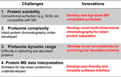
Nevertheless, the top-down MS approach still faces significant challenges in terms of protein solubility, separation, and the detection of low abundance and large proteins, as well as under-developed data analysis tools. Consequently, new technological developments are urgently needed to advance the field of top-down proteomics. We have been establishing an integrated top-down disease proteomics platform to globally examine intact proteins extracted from tissues for the identification and quantification of proteins and possible PTMs present in vivo. Specifically, we are developing novel approaches to address the current challenges in top-down MS-based proteomics.

A. To address the protein solubility challenge, we are developing new degradable surfactants that can effectively solubilize proteins and are compatible with top-down MS. we have recently developed an MS-compatible slowly degradable Surfactant (MasDeS) that can effectively solubilize proteins.24 Furthermore, we demonstrated that the solubility of membrane protein was significantly improved with the addition of this new surfactant. We are also developing different types of degradable surfactants and evaluating their performance for top-down proteomics.

B. To address the proteome complexity challenge, we are developing new chromatography materials and novel multi-dimensional liquid chromatography (MDLC) strategies to separate intact proteins. To address the proteome complexity challenge, we are developing new chromatography materials and novel strategies for multi-dimensional liquid chromatography (MDLC) to separate intact proteins. We have demonstrated the use of ultrahigh-pressure size exclusion chromatography (UHP-SEC)and hydrophobic interaction chromatography (HIC)for top-down proteomics. Moreover, we have developed a novel 3DLC strategy by coupling HIC with ion exchange chromatography (IEC) and reverse phase chromatography (RPC) for intact protein separation. We demonstrated that this 3D (IEC-HIC-RPC) approach greatly outperformed the conventional 2D IEC-RPC approach. We are now developing novel chromatography materials for intact protein separation.

C. To address the proteome dynamic range, we have been developing novel nanomaterials that can bind low abundance proteins with PTMs (e.g. phosphorylation) with high specificity in collaboration with a nanotechnologist, Prof. Song Jin (U. of Wisconsin). The current focus is to develop multivalent nanoparticle (NP) reagents for capturing phosphoproteins globally out of the human proteome for top-down MS analysis of intact phosphoproteins.

D. To address the challenge in under-developed software, we are developing user-friendly and versatile software interface for comprehensive analysis of high-resolution top-down MS-based proteomics data. Previously, we have developed a MASH Suite, a versatile and user-friendly software interface for processing, interpreting, visualizing and presenting high-resolution MS data. Recently, we have developed MASH Suite Pro, a comprehensive, user-friendly and freely available program tailored for top-down high-resolution mass spectrometry (MS)-based proteomics (Manuscript submitted). MASH Suite Pro significantly simplifies and speeds up the processing and analysis of top-down proteomics data by combining tools for protein identification, quantitation, characterization, and validation into a customizable and user-friendly interface.
We envision that by taking this multi-pronged approach to overcome the challenges facing top-down proteomics, we will significantly advance the burgeoning top-down proteomics field, which recently gained momentum through the creation of the Consortium for Top-down Proteomics


Friday, 13 July 2018

Systems proteomics of liver mitochondria function


Expanded proteomic analysis of metabolism


Combined analysis of large data sets characterizing genes, transcripts, and proteins can elucidate biological functions and disease processes. Williams et al. report an exceptionally detailed characterization of mitochondrial function in a genetic reference panel of recombinant inbred mice. They measured the metabolic function of nearly 400 mice under various environmental conditions and collected detailed quantitative information from livers of the animals on over 25,000 transcripts. These data were integrated with quantitation of over 2500 proteins and nearly 1000 metabolites. Such analysis showed a frequent lack of correlation of transcript and protein abundance, enabled the identification of genomic variants of mitochondrial enzymes that caused inborn errors in metabolism, and revealed two genes that appear to function in cholesterol metabolism.

Structured Abstract

INTRODUCTION

Over the past two decades, continuous improvements in “omics” technologies have driven an ever-greater capacity to define the relationships between genetics, molecular pathways, and overall phenotypes. Despite this progress, the majority of genetic factors influencing complex traits remain unknown. This is exemplified by mitochondrial supercomplex assembly, a critical component of the electron transport chain, which remains poorly characterized. Recent advances in mass spectrometry have expanded the scope and reliability of proteomics and metabolomics measurements. These tools are now capable of identifying thousands of factors driving diverse molecular pathways, their mechanisms, and consequent phenotypes and thus substantially contribute toward the understanding of complex systems.

RATIONALE

Genome-wide association studies (GWAS) have revealed many causal loci associated with specific phenotypes, yet the identification of such genetic variants has been generally insufficient to elucidate the molecular mechanisms linking these genetic variants with specific phenotypes. A multitude of control mechanisms differentially affect the cellular concentrations of different classes of biomolecules. Therefore, the identification of the causal mechanisms underlying complex trait variation requires quantitative and comprehensive measurements of multiple layers of data—principally of transcripts, proteins, and metabolites and the integration of the resulting data. Recent technological developments now support such multiple layers of measurements with a high degree of reproducibility across diverse sample or patient cohorts. In this study, we applied a multilayered approach to analyze metabolic phenotypes associated with mitochondrial metabolism.

RESULTS

We profiled metabolic fitness in 386 individuals from 80 cohorts of the BXD mouse genetic reference population across two environmental states. Specifically, this extensive phenotyping program included the analysis of metabolism, mitochondrial function, and cardiovascular function. To understand the variation in these phenotypes, we quantified multiple, detailed layers of systems-scale measurements in the livers of the entire population: the transcriptome (25,136 transcripts), proteome (2622 proteins), and metabolome (981 metabolites). Together with full genomic coverage of the BXDs, these layers provide a comprehensive view on overall variances induced by genetics and environment regarding metabolic activity and mitochondrial function in the BXDs. Among the 2600 transcript-protein pairs identified, 85% of observed quantitative trait loci uniquely influenced either the transcript or protein level. The transomic integration of molecular data established multiple causal links between genotype and phenotype that could not be characterized by any individual data set. Examples include the link between D2HGDH protein and the metabolite D-2-hydroxyglutarate, the BCKDHA protein mapping to the gene Bckdhb, the identification of two isoforms of ECI2, and mapping mitochondrial supercomplex assembly to the protein COX7A2L. These respectively measured variants in these mitochondrial proteins were in turn associated with varied complex metabolic phenotypes, such as heart rate, cholesterol synthesis, and branched-chain amino acid metabolism. Of note, our transomics approach clarified the contested role of COX7A2L in mitochondrial supercomplex formation and identified and validated Echdc1 and Mmab as involved in the cholesterol pathway.

CONCLUSION

Overall, these findings indicate that data generated by next-generation proteomics and metabolomics techniques have reached a quality and scope to complement transcriptomics, genomics, and phenomics for transomic analyses of complex traits. Using mitochondria as a case in point, we show that the integrated analysis of these systems provides more insights into the emergence of the observed phenotypes than any layer can by itself, highlighting the complementarity of a multilayered approach. The increasing implementation of these omics technologies as complements, rather than as replacements, will together move us forward in the integrative analysis of complex traits.

Wednesday, 11 July 2018

Clinical Chemistry


Abstract


BACKGROUND: There is an urgent need for blood-based molecular tests to assist in the detection and diagnosis of cancers at an early stage, when curative interventions are still possible, and to predict and monitor response to treatment and disease recurrence. The rich content of proteins in the blood that are impacted by tumor development and host factors provides an ideal opportunity to develop noninvasive diagnostics for cancer.


CONTENT: Mass spectrometry instrumentation has advanced sufficiently to allow the discovery of protein alterations directly in plasma across no less than 7 orders of magnitude of protein abundance. Moreover, the use of proteomics to harness the immune response in the form of seropositivity to tumor antigens has the potential to complement circulating protein biomarker panels for cancer detection. The depth of analysis currently possible in a discovery setting allows the detection of potential markers at concentrations of less than 1 μg/L. Such low concentrations may exceed the limits of detection of ELISAs and thus require the development of clinical assays with exquisite analytical sensitivity. Clearly, the availability for discovery and validation of biospecimens that are highly relevant to the intended clinical application and have been collected, processed, and stored with the use of standard operating procedures is of crucial importance to the successful application of proteomics to the development of blood-based tests for cancer.


SUMMARY: The realization of the potential of proteomics to yield blood biomarkers will benefit from a collaborative approach and a substantial investment in resources.

For disease investigation, the profiling of blood constituents, notably serum and plasma, using protein characterization technologies holds long-standing interest because of the easy accessibility of this circulating fluid and its rich content of proteins that inform scientists about the health status of an individual. The available methodologies to analyze proteins have evolved dramatically over the past few decades. The initial method consisted of 1-dimensional protein separations, which was followed by the use of 2-dimensional polyacrylamide gel electrophoresis coupled with Edman sequencing. The advent of mass spectrometry, coupled with the sequencing of human and other genomes, has had a dramatic impact on the field of proteomics. The capabilities of current proteomics technologies in terms of coverage of the proteome and depth of analysis that can be achieved in a quantitative manner are truly astounding compared with just a decade ago. Recent advances include substantial increases in speed, analytical sensitivity, and dynamic range, and the availability of multiple fragmentation techniques. Equally important is orthogonal sample fractionation before mass spectrometry. Yet there remains a perception that proteomics technologies are inadequate to address the protein complexities inherent in cells, tissues, and biological fluids. Here we outline strategies for the application of proteomics to the development of blood-based cancer markers.

Reference: http://clinchem.aaccjnls.org/content/59/1/119

Protein Identification


Proteomics is the study of the complete set of proteins that is expressed at a given time in a cell, tissue, organ or organism, i.e. the proteome. Proteins are the machinery of the cell, responsible for performing the functions essential for cells to operate, survive, grow and divide. Proteins are also e.g. responsible for relaying signals or messages within and between cells that drive biological and metabolic processes. In addition, proteins are the main constituent of the physical structures of cells and organism.
There is a general interest to proteomics in life sciences since as compared to e.g. genomics/functional genomics, proteomics is able to give a much more complete understanding of the studied biological system. Due to events like alternative splicing and post-translational modifications (like phosphorylation, glycosylation, ubiquitinylation, myristyolation, palmityolation etc.), the number of different proteins expressed in an organism is by far greater than the number of its functional genes. The expression level of functional proteins is regulated at many post-transcriptional levels, like the action of inhibitory RNA species, protein localization and degradation, in addition, post-translational modifications, like phosphorylations, can have the role of switching an inactive protein to active. Due to this reasons, simple mRNA transcript measurements by functional genetics may give a biased reading of the actual protein activity.

In most cases, changes at protein level determine the differences between sickness and health and can be used to monitor the progression of a disease. Almost all drugs on the market and being developed act by trying to alter a protein (mal)function. Therefore, typically one of the first steps in the drug discovery project is the identification and validation of the protein target(s) or markers associated with a disease (one type of biomarkers).
Our expertise in proteomics

We offer our customers a wide spectrum of proteomics expertise and services. Our services are based on nanoLC-MS/MS and other instrumentation platforms, coupled with bioinformatics and computational systems biology. The samples can be fluids, like serum, plasma, urine, cell lysates or solid tissue samples as well as processed samples like SDS gel plugs.

Monday, 9 July 2018

Microbial Proteomics

Microbes are by far the most abundant organisms living on our planet, outnumbering the human population by more than 1020 times. Even our own body is populated by 10 times more microbial than human cells. In the light of such numbers it becomes apparent, that we will always have to deal with all microbial life forms that surround us, and that we have to share living space and resources. Therefore, a thorough understanding of microbial physiologies, infection and defense systems, metabolic processes or survival strategies are of great interest in order to actively defeat or cultivate microorganism.

Our group works on microorganisms, on the one hand as model organisms and for technology development, on the other hand because of their clinical relevance. We currently focus on Saccharomyces cerevisiae, Mycobacterium tuberculosis, Streptococcus pyogenes, as well as various viruses. In recent years, for most of these microbes we have generated comprehensive, high quality assay repositories for the sensitive detection and relative as well as absolute quantification of virtually all annotated proteins using targeted mass spectrometric techniques like SRM or SWATH-MS.
In the context of M. tuberculosis we use these unique resources to improve genome annotation by proteogenomic analysis, as well as to study protein-level responses to different stress conditions, such as starvation and oxygen depletion, which causes a transition into a clinically relevant dormant state. Further, we are interested in elucidating host-pathogen interactions upon infection of human cells, such as altered protein expression in M. tuberculosis, as well as changes in the human MHC peptidome.

In the case of S. pyogenes our aim is to identify and understand virulence factors by integrating genomic, proteomic, and phenotypic data of clinically isolated S. pyogenes strains. Specifically, we aim to understand how genomic variation may lead to differences in proteome expression and ultimately differences in phenotype (virulence).

Our research interests in the smallest eukaryotic model organism S. cerevisiae are multifaceted. One focus is on the better understanding of metabolic regulation and adaptation upon changing nutritional conditions based on protein abundance changes as well as phosphoproteomic analyses. Further, we are interested in the phosphorylation-based signaling networks, including for example the TOR pathway and the pheromone pathway. Also the regulation of gene expression by transcriptional regulatory elements we investigate using targeted mass spectrometry. Finally, we use yeast as a model organism to study how genetic loci are influencing the levels of related proteins by quantitative trait loci analysis (QTLs).


Reference; http://www.imsb.ethz.ch/research/aebersold/research/microbial-proteomics.html 
New rendering from 3D Protein Imaging. Reproduction of the current molecule of the month https://lnkd.in/gFJ7sCR made with the new version of the Protien Imager that will be soon available at https://lnkd.in/dCb2u66 #proteinimaging #molecularbiology #molecularmodeling #molecularneuroscience #molecularart #3dproteinimaging #rendering #reproduction #3d

Friday, 6 July 2018

Proteomics Cells

Researchers can count on improved proteomics method: 


Every cell in the body contains thousands of different protein molecules and they can change this composition whenever they are induced to perform a particular task or convert into a different cell type. Understanding how cells function depends on proteomics, the ability to measure all of the changes in a cell's protein components.
In a recent paper published in the journal Analytical Chemistry, Martin Wühr and colleagues in Princeton University's Department of Molecular Biology described an improved method to accurately count the proteins present in a cell under different circumstances.

The basic tool for counting proteins is a machine called a mass spectrometer. Cell samples can be run through this type of instrument one at a time, but this is laborious and it can be difficult to detect any changes between different samples. An alternative approach is to label all of the proteins in a particular sample with a unique "isobaric" tag. Multiple samples--up to 11--can then be mixed together and run through the mass spectrometer at the same time, with the isobaric tag functioning as an identifying barcode that tells the researcher which sample the protein originally came from. This speeds things up and makes it easier to quantify any changes in the protein composition of different samples.
"However, with the simplest version of isobaric tagging, known as TMT-MS2, there are major difficulties in distinguishing real signals from background noise," Wühr explains. "That makes the readouts unreliable and only semi-quantitative."
A more complex version of isobaric tagging, called TMT-MS3, can improve this signal-to-noise problem, but it is slower and less sensitive. Moreover, it relies on a much more expensive type of mass spectrometer beyond the reach of most researchers.
While he was a postdoc at Harvard University, Wühr developed a different approach to isobaric tagging that solved the signal-to-noise problem while remaining compatible with cheaper, widely available mass spectrometers. But the technique--known as TMTc--was not without its own problems, particularly a lack of precision that made it hard to obtain consistent results.
In their recent Analytical Chemistry paper, Wühr and two of his graduate students, Matthew Sonnett and Eyan Yeung, described an improved version of TMTc that they named TMTc+. By changing how the cell samples are prepared and altering the computer algorithm that extracts data from the mass spectrometer, Wühr and colleagues were able to address many of the limitations associated with the various methods of isobaric tagging.
"The TMTc+ method is in a kind of sweet spot compared to the other methods," Wühr says. "It provides superb measurement accuracy and precision, it's at least as sensitive as any other method, and it's compatible with around ten times more mass spectrometers than TMT-MS3."
Naturally, Wühr says, there is still room for improvement. TMTc+ only allows a maximum of 5 samples to be run at the same time, and the detection of proteins in these samples is relatively inefficient. Both of these problems can be solved by developing new types of isobaric tags. "We have to explore the chemical space of these tags and find ones that work really well," Wühr says. "To this end, we have started a collaboration with the Carell group, organic chemistry experts at the LMU Munich, and already published a proof of principle paper. Eventually, these efforts should lead to an approach that will allow researchers to count every protein in a cell as it changes its form and function."

Host/Pathogen Proteomics

Host/Pathogen Proteomics: Mycoplasma pneumoniae

The bacterium Mycoplasma pneumoniae colonizes host pulmonary epithelium and is the most common cause of human community-acquired pneumonia. It successfully avoids detection by the host immune system, as the microbe alters its own cell membrane to mimic its host in order to establish chronic respiratory infections. Progression to autoimmune disease is not unknown.

Initial infection by M. pneumoniae stimulates production and release of proteins by the host cell in response to binding to the infectious agent and its internalization. Li et al. (2014) used a label-free shotgun quantitative proteomics approach to investigate the host secretome.1By characterizing the biologically active proteins released in this initial phase, the researchers hope to elucidate the pathways involved and the roles of these secretory proteins in disease pathogenesis.

Li and colleagues used the human alveolar carcinoma cell line A549 to establish initial proteomic events following infection by M. pneumoniae. They chose airway epithelial cells because these are the most common primary cells colonized by the microorganism upon infection.


The researchers cultured A549 cells either with or without the infectious agent before harvesting the conditioned media. The scientists analyzed the tryptic digests by nano liquid chromatography–tandem mass spectrometry (LC-MS/MS) using an LTQ Velos ion trap mass spectrometer (Thermo Scientific). The authors searched the data obtained against the IPI human protein database v3.60 to identify proteins secreted by the cell cultures.

Initial LC-MS/MS quantification using DeCyder software showed that 113 out of the 256 proteins identified showed at least 1.5-fold differential expression between the control (uninfected) and the infected cell cultures. Of these, 65 were elevated in abundance and 48 were reduced in the infected cells. Nine proteins were found only in the uninfected control cells, whereas 10 were exclusive to the cells post-infection. Interleukin-33 (IL-33) was one of these proteins, and the researchers confirmed increased levels following infection by utilizing an enzyme-linked immunosorbent assay (ELISA).

The researchers confirmed the proteomics results using Western blotting and real-time polymerase chain reaction (PCR) for selected proteins. Immunoblotting identified the same proteins in cell lysates and conditioned media, showing results consistent with the LC-MS/MS findings for proteins ADAM9, SERPINE1, IL-33, IGFBP4, Gal-1, and MIF, which were more abundant in conditioned media from infected cells.

Out of the 256 identified, over 59% (n = 152) of the proteins were classified as either classical secretory (n = 83) or non-classical secretory (n = 69). A total of 190 proteins were associated with exosomes. Further analysis using GO classification showed that the majority were nuclear-associated. The researchers used DAVID 6.7 for functional annotation clustering analysis and found that most proteins were associated with vesicles or were from the extracellular region or matrix.

Using the KEGG database for pathway analysis, the researchers found that those associated with metabolism, infection and proliferation were over-represented in the proteins identified post-infection. Li and co-workers further analyzed their data to discover more about the functional processes targeted by the post-infection secretome, using the BiNGO tool and STRING algorithm to examine differentially expressed proteins. The STRING analysis highlighted clusters involving stress and immune response pathways, among others. In summary, pathway analyses gave the researchers indications of where M. pneumoniae could induce protein secretion that alters the host cell function.

Finally, Li and co-authors repeated their investigation and found similar proteins in broncheoaveolar lavage samples and plasma from patients with confirmed M. pneumoniae infection. When they measured IL-33 in these two samples, they found elevated levels compared with a control group of patients who presented with respiratory foreign body. Statistical testing showed that IL-33 could indeed be used as a diagnostic marker of infection.

Overall, Li et al. are confident that, by characterizing the post-infection secretome, they have uncovered new regulatory pathways in the host response to M. pneumoniae that can be explored further to elucidate disease pathogenesis and treatment options.

Reference: https://www.thermofisher.com/blog/proteomics/hostpathogen-proteomics-mycoplasma-pneumoniae/

Monday, 2 July 2018

Proteomics- Methodology for Studying RNA/Protein interactions

Protein microarray is one of the technology and it is a critical tool in biochemistry and molecular biology. The analytical microarray is the most powerful multiplexed detection technology. The functional microarray is one of the important tools for the high-throughput and large-scale system for biological studies. Some application of functional microarray is the detection of protein binding properties as protein-protein interaction, protein -DNA interaction, protein-RNA interaction and antigen-antibody interaction. There are numerous cellular processes in which protein kinases are involved. Recording and analyzing of immune responses.

An atlas of the aging lung mapped by single cell transcriptomics and deep tissue proteomics


Abstract


Aging promotes lung function decline and susceptibility to chronic lung diseases, which are the third leading cause of death worldwide. We used single-cell transcriptomics and mass spectrometry to quantify changes in cellular activity states of 30 cell types and the tissue proteome from lungs of young and old mice. Aging led to increased transcriptional noise, indicating deregulated epigenetic control. We observed highly distinct effects of aging on cell type level, uncovering increased cholesterol biosynthesis in type-2 pneumocytes and lipofibroblasts as a novel hallmark of lung aging. Proteomic profiling revealed extracellular matrix remodeling in old mice, including increased collagen IV and XVI and decreased Fraser syndrome complex proteins and Collagen XIV. Computational integration of the aging proteome and single cell transcriptomes predicted the cellular source of regulated proteins and created a first unbiased reference of the aging lung. The lung aging atlas can be accessed via an interactive user-friendly web tool at  https://theislab.github.io/LungAgingAtlas